- Blog /
- August 11, 2021
Everything You Need to Know about Breast CT
Breast cancer is a serious widespread disease that affects women globally. In 2020 alone, there were 2.3 million diagnosed cases around the world, out of which 685,000 proved to be fatal. In the United States, around 1 out of 8 women is expected to develop invasive breast cancer during her lifetime.
Medical professionals diagnose the condition via a breast ultrasound, MRI, biopsy, and diagnostic mammogram. Mammography has long been used and recognized as an effective tool to identify cancer symptoms early on. However, its accuracy is still not suitable for early detection and it comes with its own set of limitations. Especially for women with dense breasts, mammograms are not sensitive enough to find small lesions and often fail to provide an accurate diagnosis. An alternative to a mammogram is a breast CT scan, a technology that overcomes the shortcomings of mammography. Research suggests that breast CT is considerably better than 2D mammography as it better detects masses that have the potential of becoming malignant (P=.006).
What is a Breast CT?
A breast computerized tomography scan (CT scan) is an X-ray-based method. It provides doctors with the information they need about the breast via multiple cross-sections. CT scans have become a popular technique for evaluating organs throughout the body. This technique is especially beneficial for bigger-breasted women to help them detect the extent or spread of a malignant tumor and to figure out if a mastectomy can help remove it. Breast CT scans show signs of primary cancer and that of any local and distant nodal metastases that might be present.
Breast CT scans can reduce the substantial discomfort that mammography and other cancer detection methods can cause to patients. Moreover, this type of imaging eliminates any tissue overlap and effectively detects any soft tissue lesions. Breast CT images are also produced in a shorter amount of time than those produced by conventional mammography.
Breast CT requires no breast compression, and it creates an image that doctors can view and reconstruct in any plane. Thus, doctors get a full view of the breast without needing to send the patient to additional exams; Medical professionals can view 3D reconstructed images in axial, coronal, and sagittal planes.
What Happens During a Breast CT Scan?
Once you are changed and prepped for the CT scan, often doctors will administrate a contrast medium so that images appear as clear as possible.
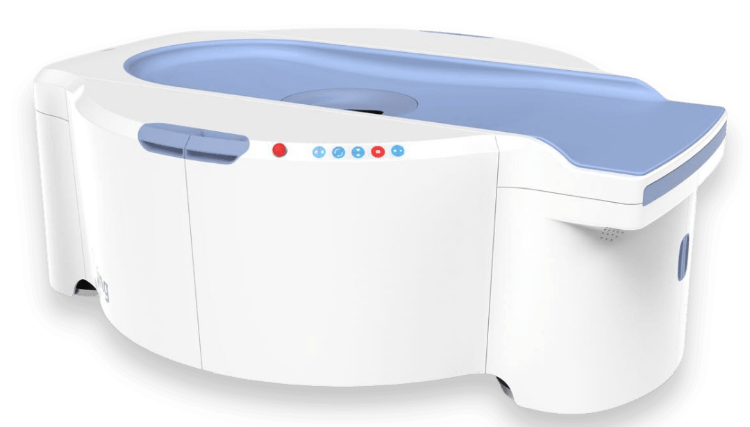
For the scan itself, you will lie down in a prone position while a radiographer monitors you from outside the room. The machine will then take pictures while producing a whirring noise. This process usually lasts for a few seconds while images are produced.
Breast CT is a safe cancer detection method for most people. All in all, 3D breast CT technology offers superior visualization and diagnostic analysis of cell and tissue abnormalities, even in dense breasts. The radiation involved is similar to that of conventional mammograms, and the overall process is much more comfortable for patients.
Status of Breast CT in Today’s Breast Imaging Environment
The American Medical Association recently added six new Breast CT CPT codes to allow for clinic reimbursement. Reimbursement has already been accepted by private payors and Medicare.
Talk to your referring physician about getting a breast CT scan as your annual breast exam.
Sources:
https://www.ncbi.nlm.nih.gov/pmc/articles/PMC4201870/
https://www.breastcancer.org/symptoms/testing/types/cat_scans#
